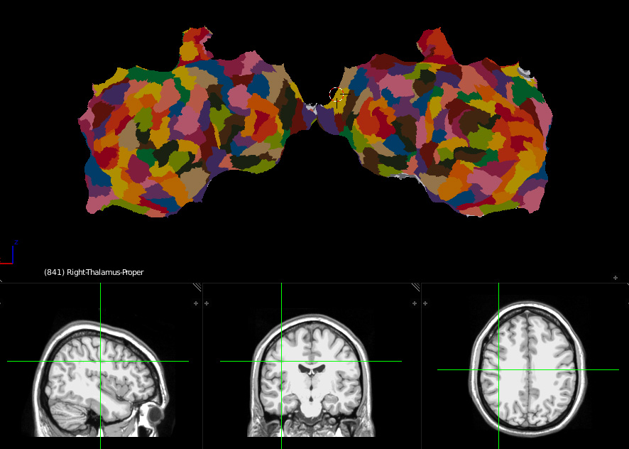
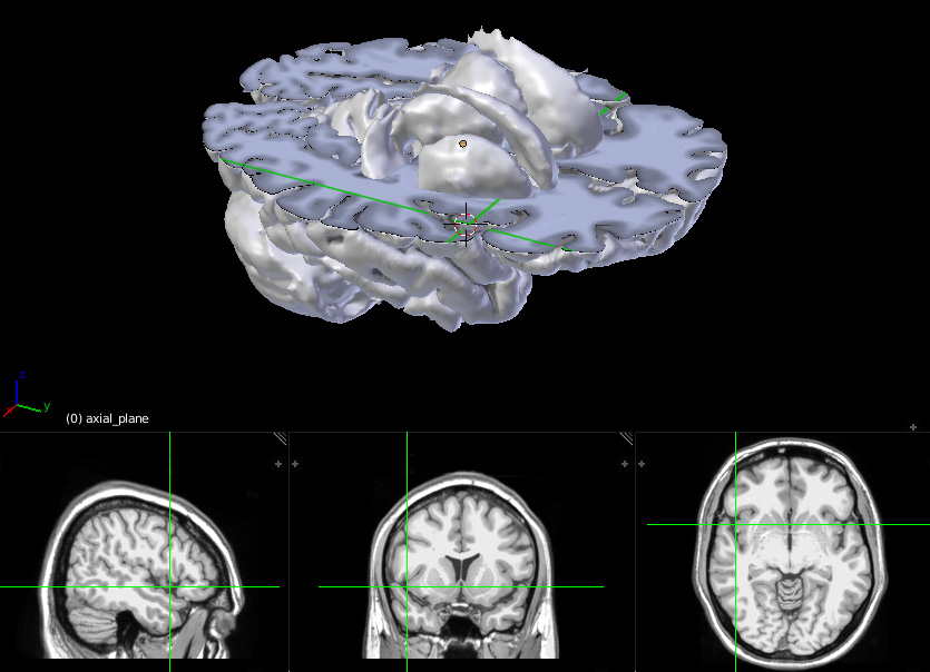
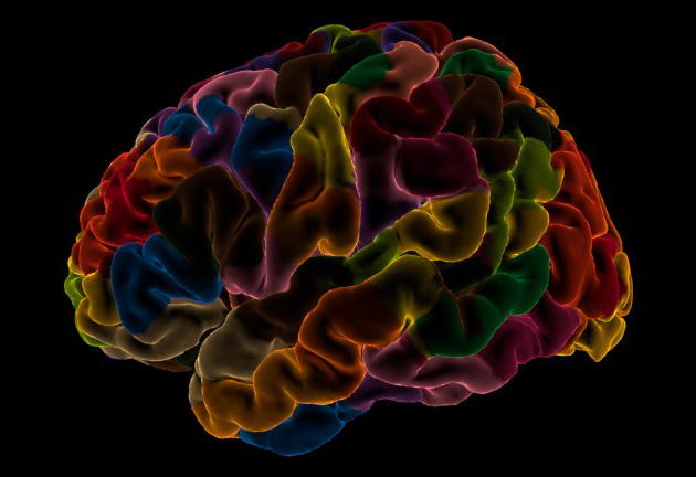
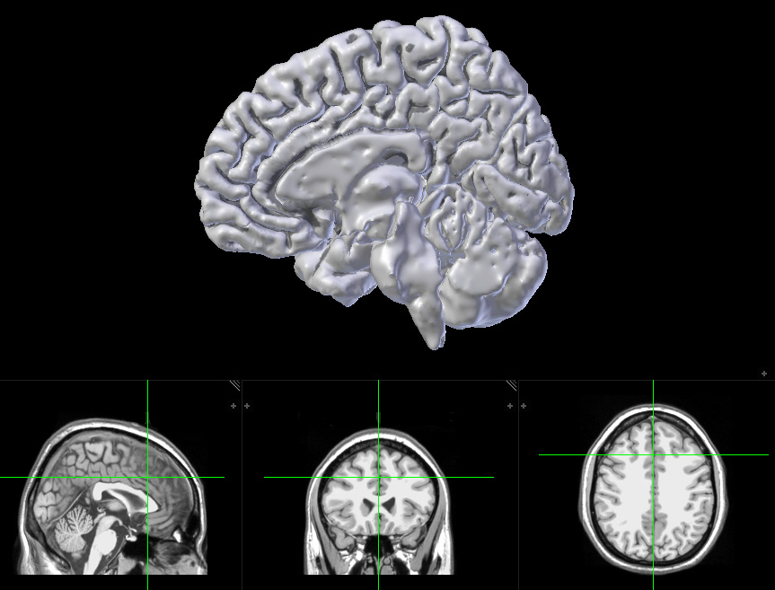
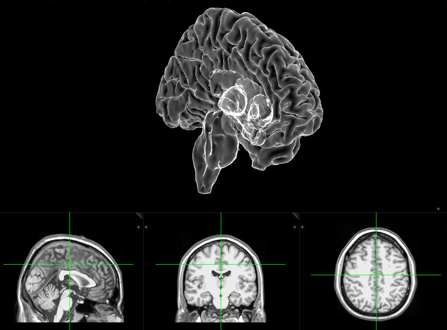
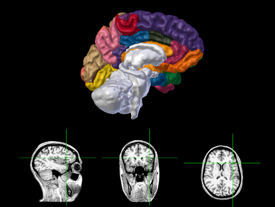
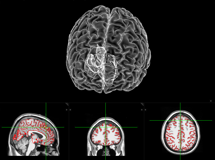
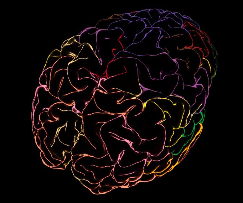
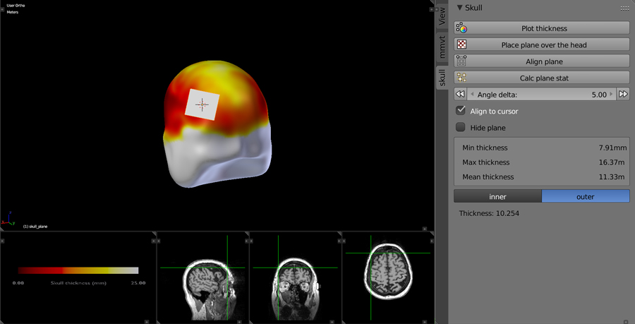
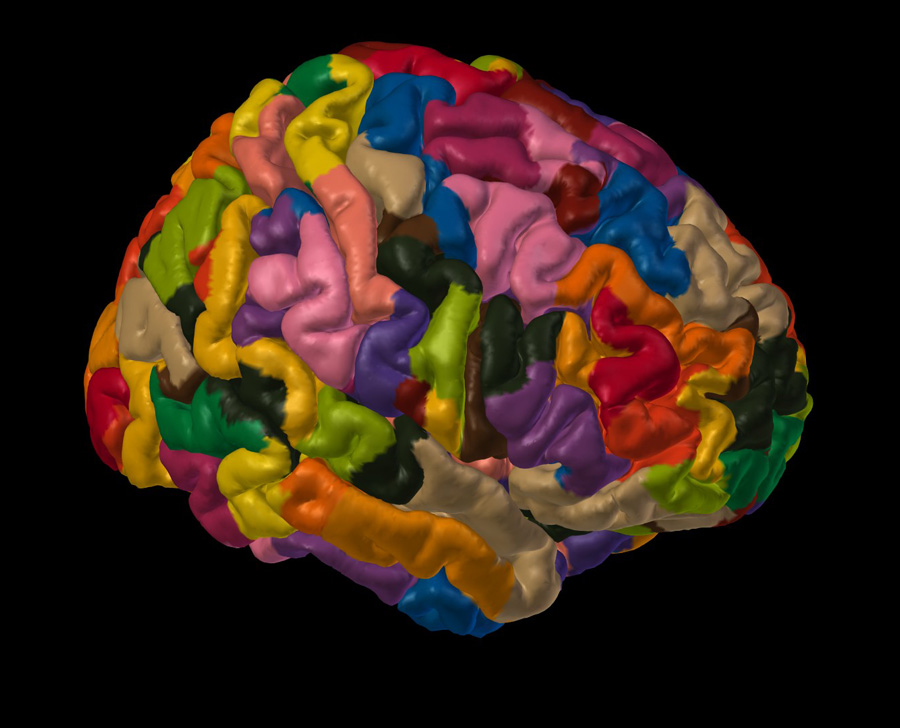
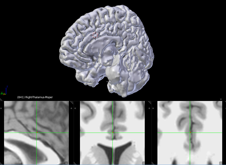
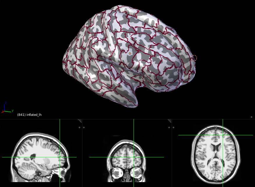
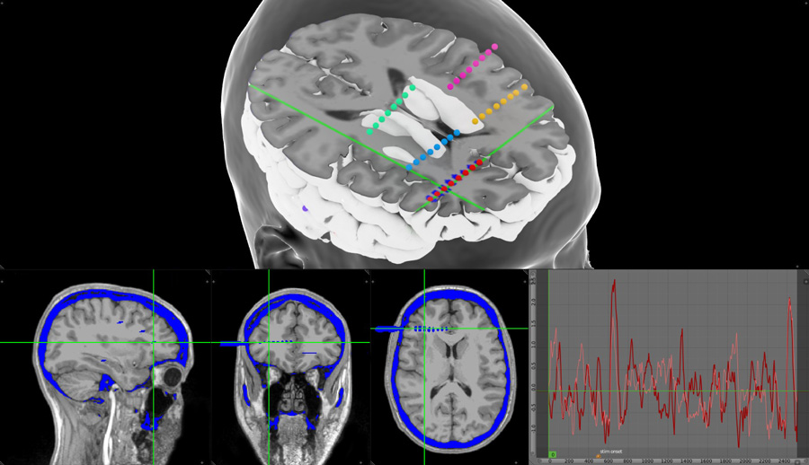
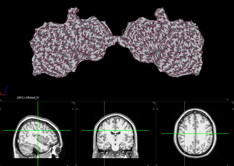
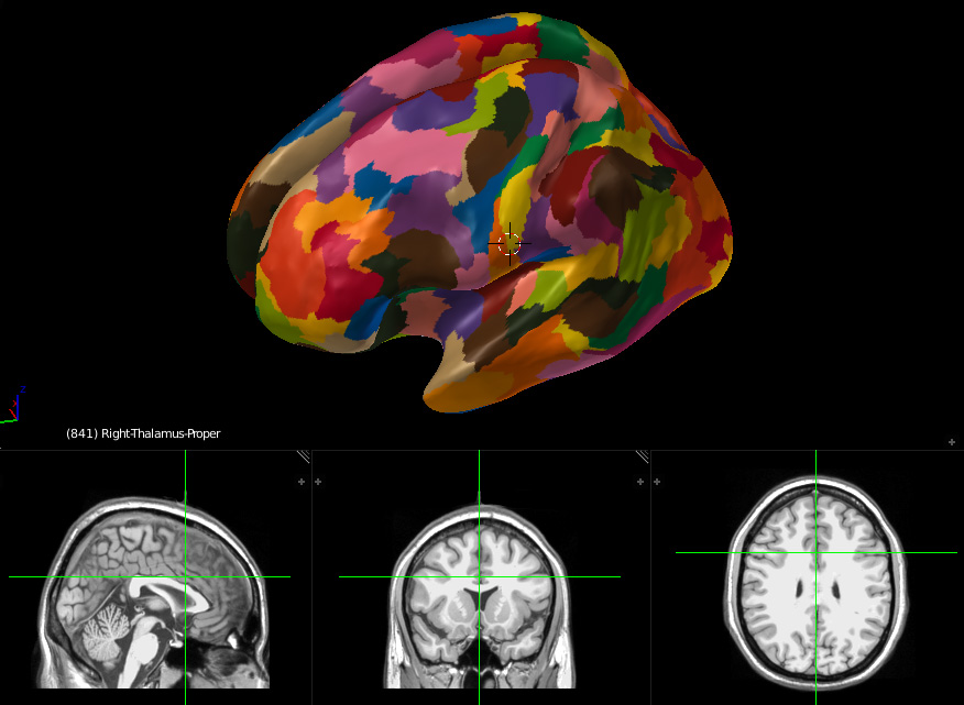
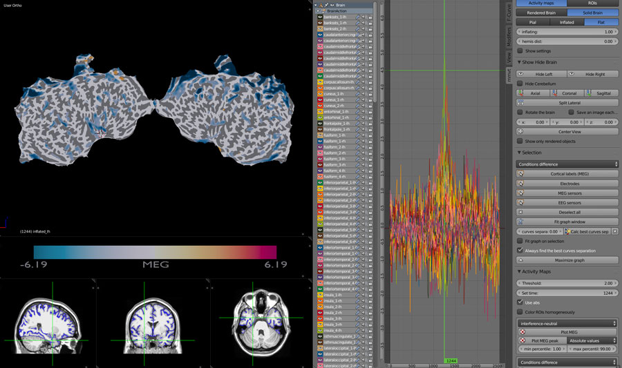
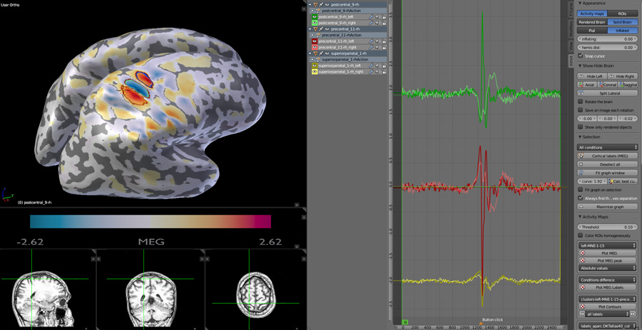
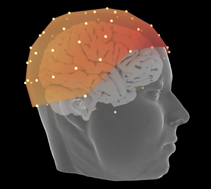
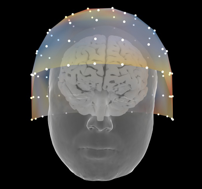
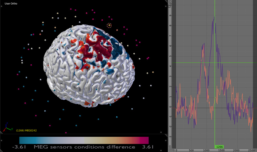
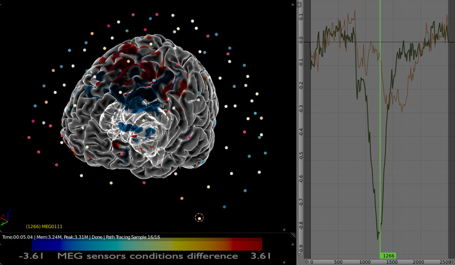
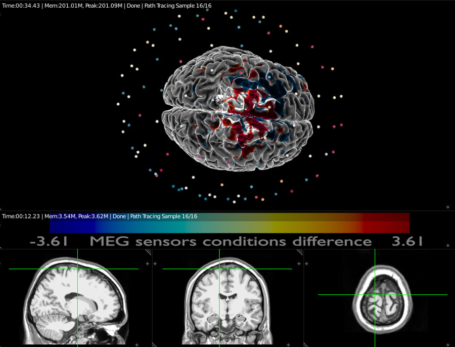
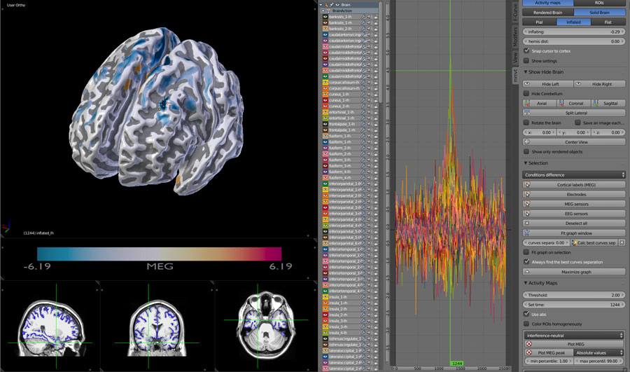
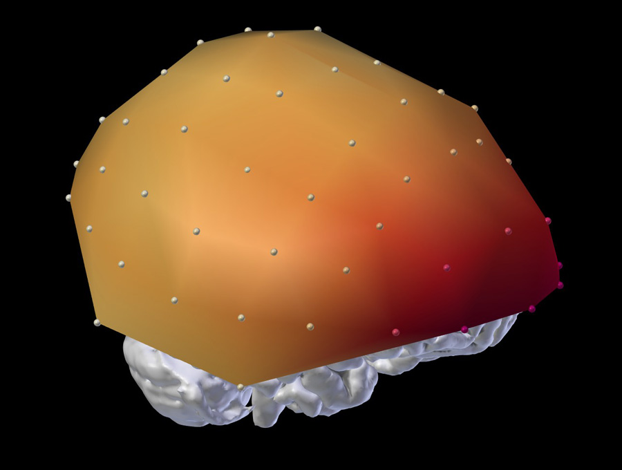
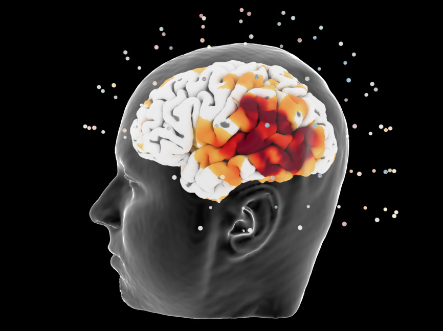
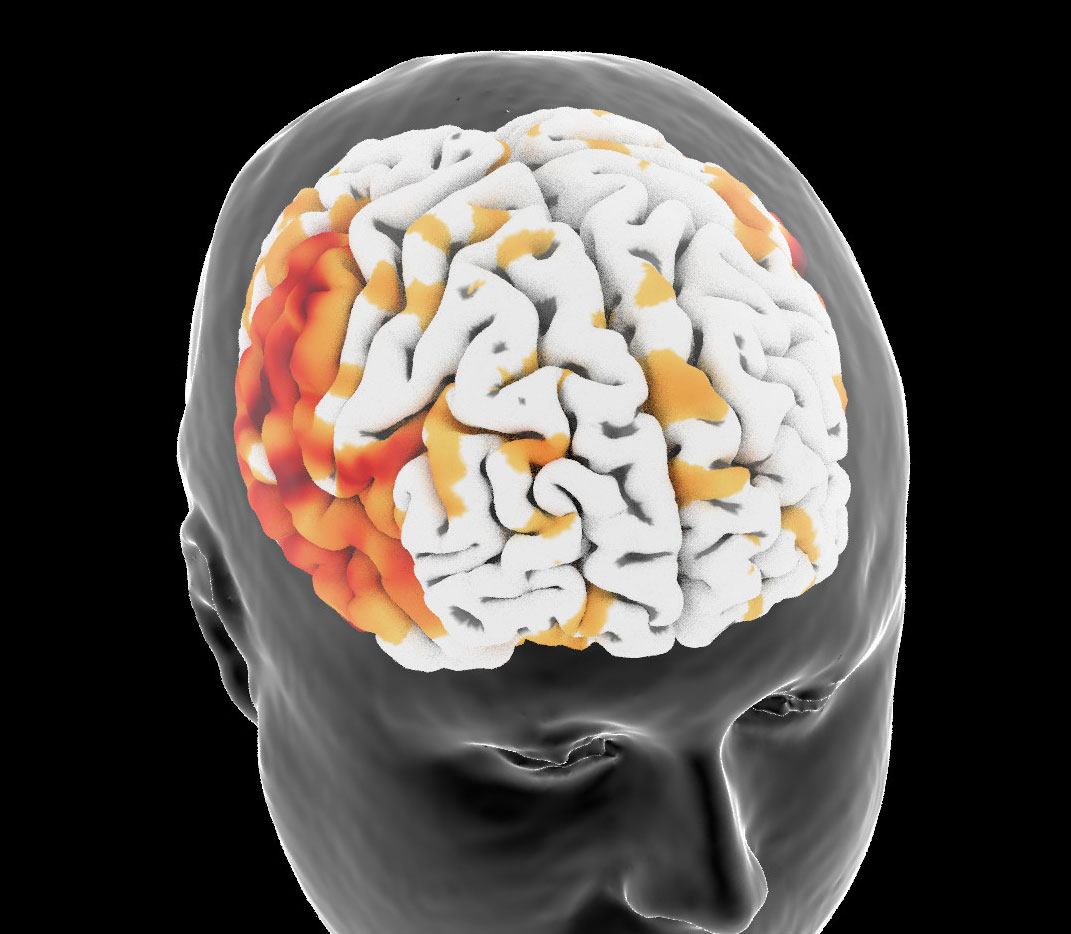
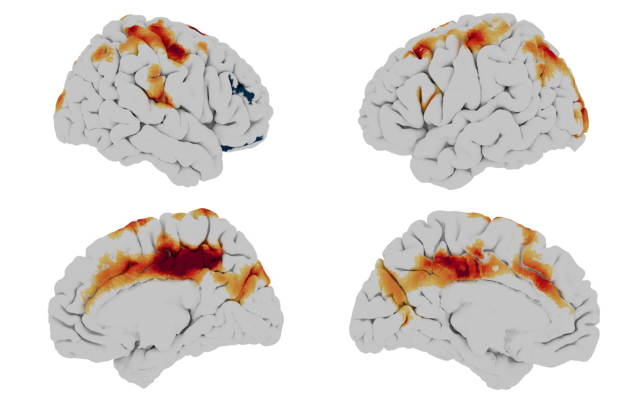
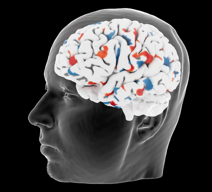
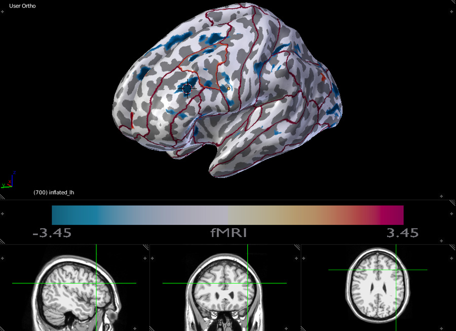
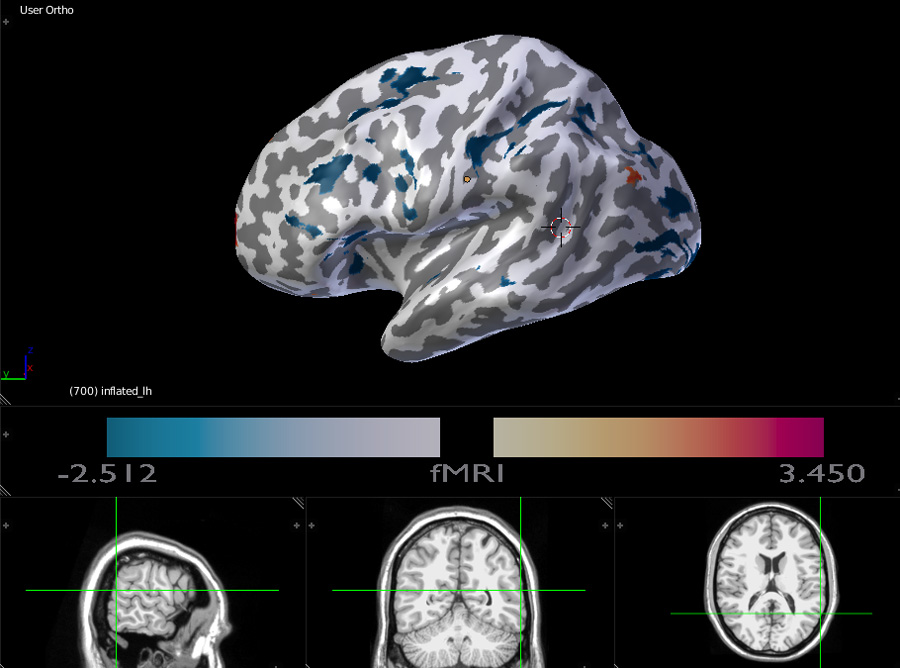
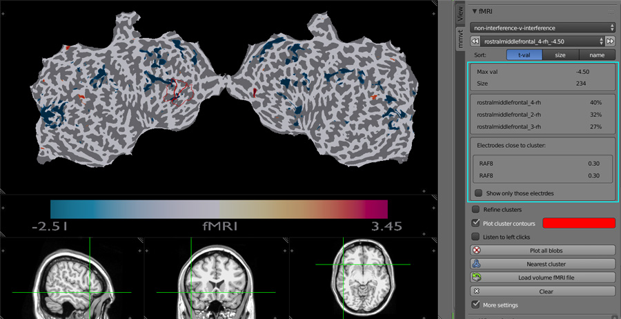
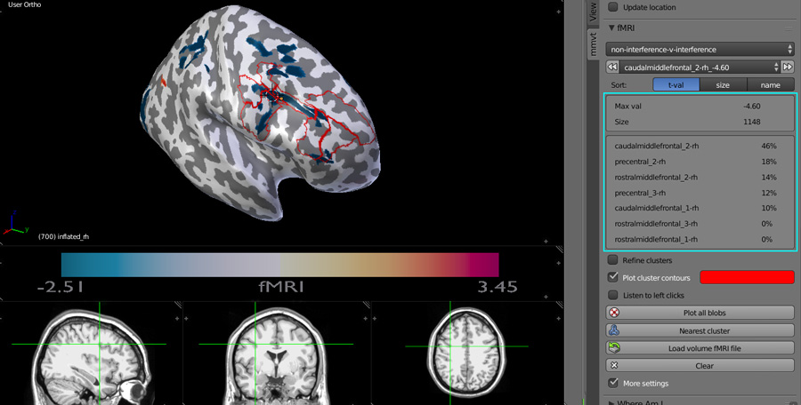
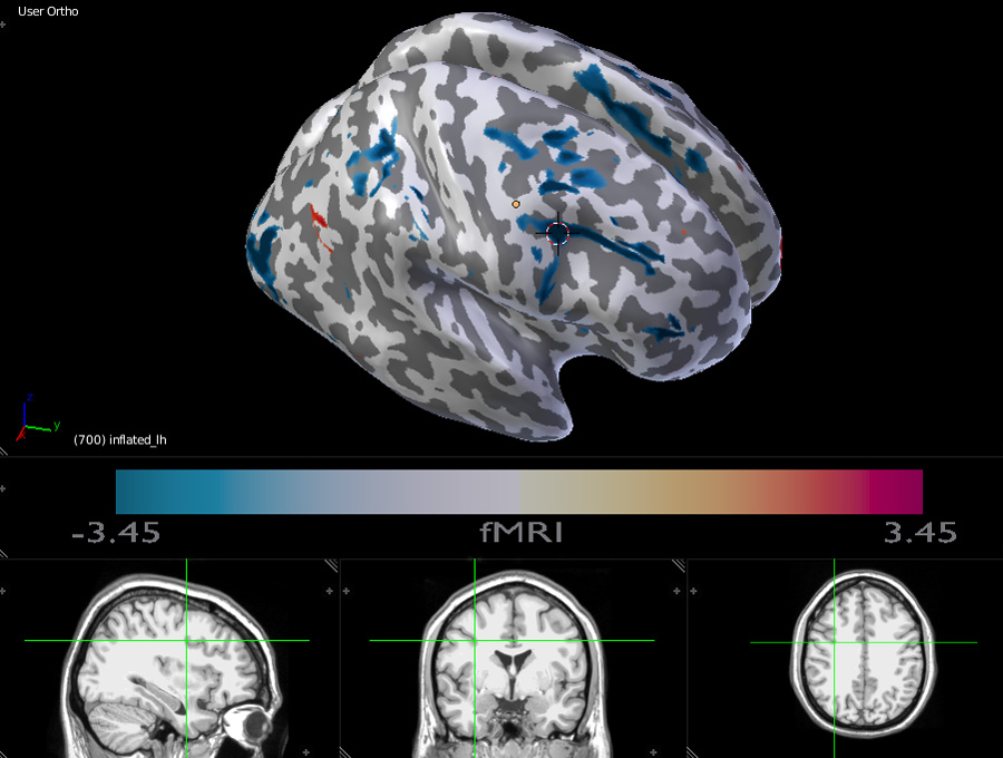
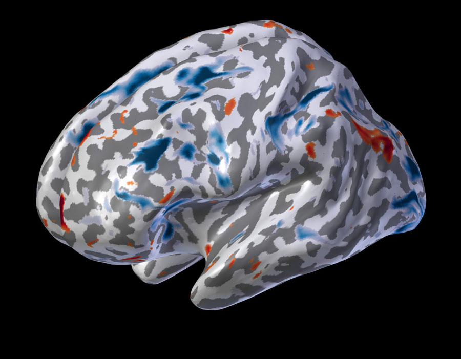
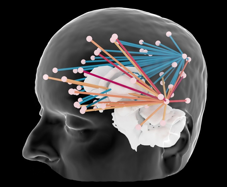
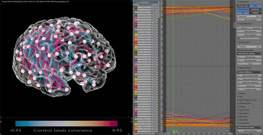
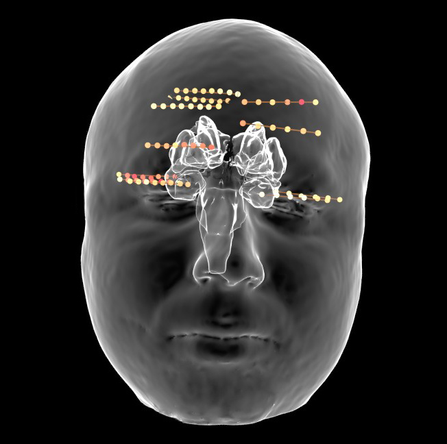
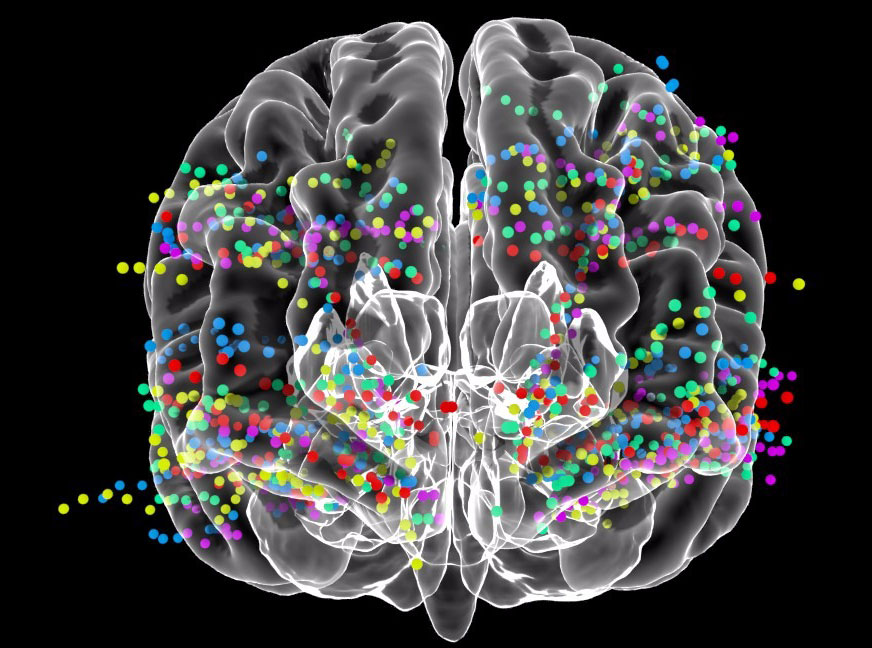
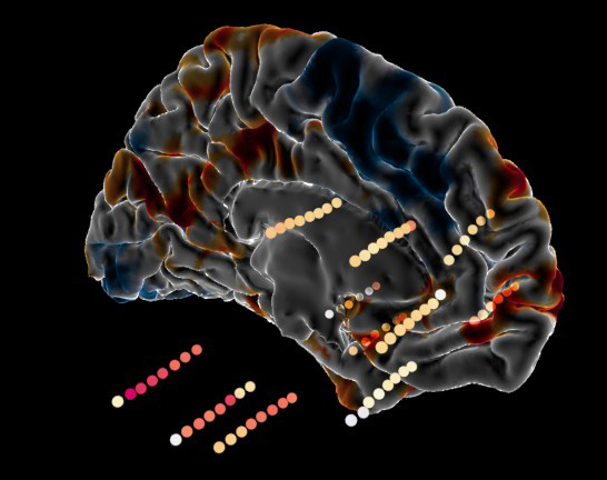
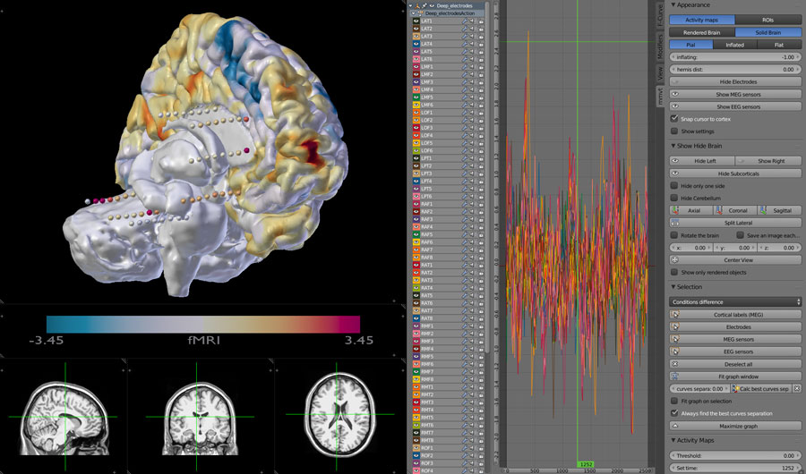
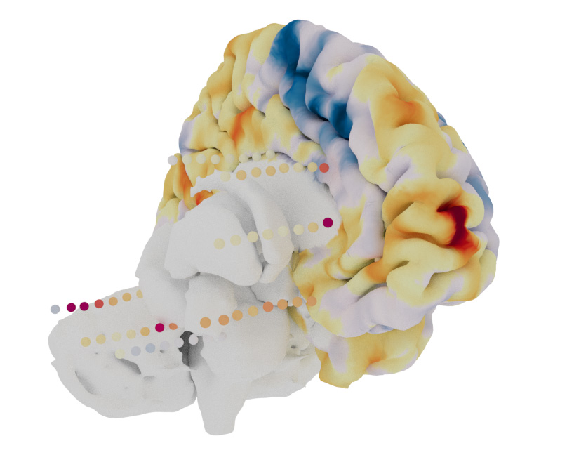
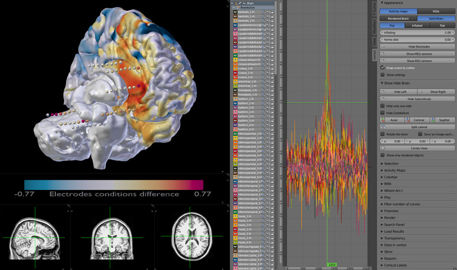
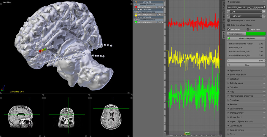
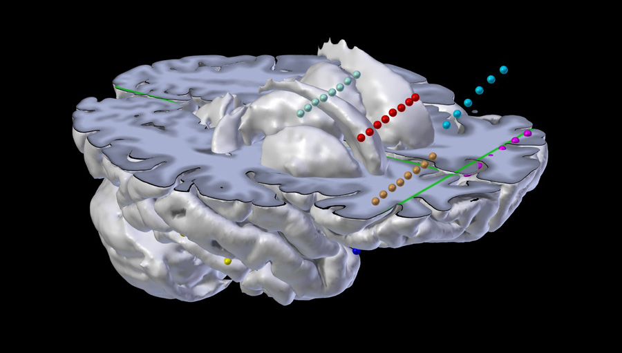
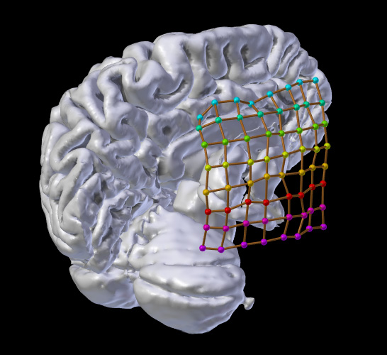
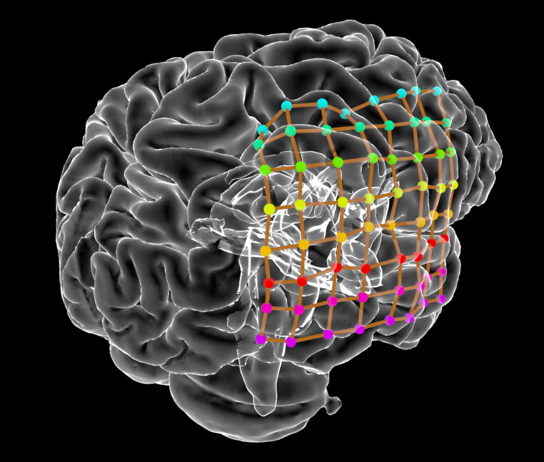
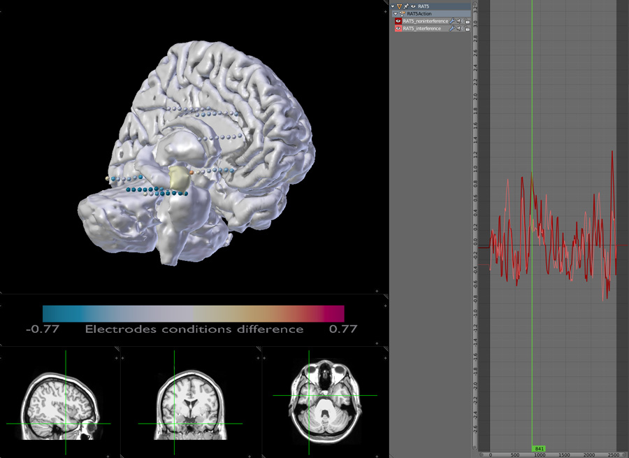
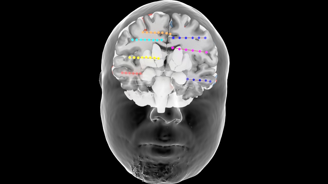
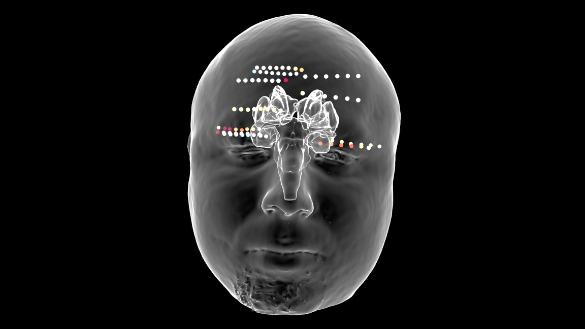
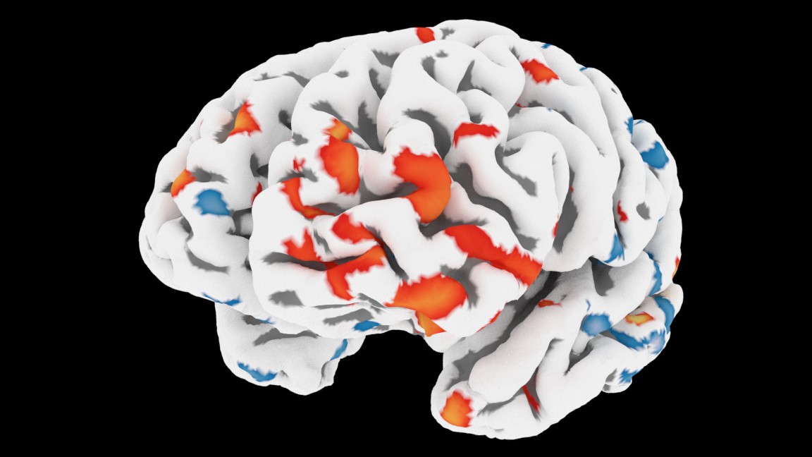
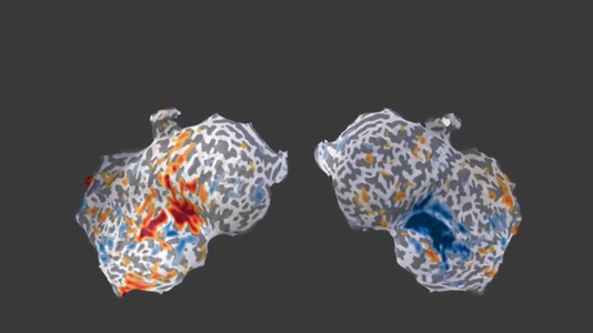
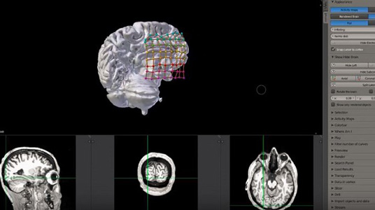
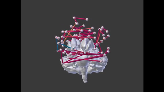
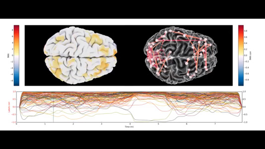
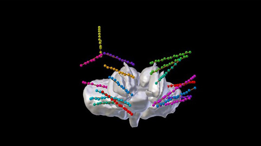
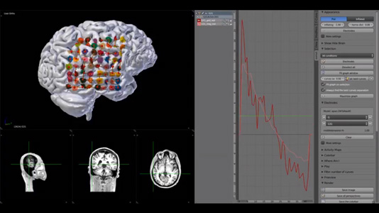
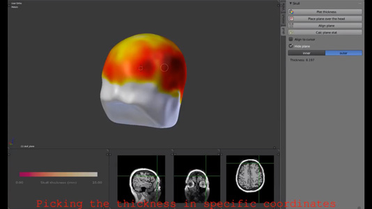
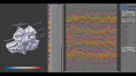
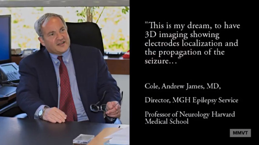
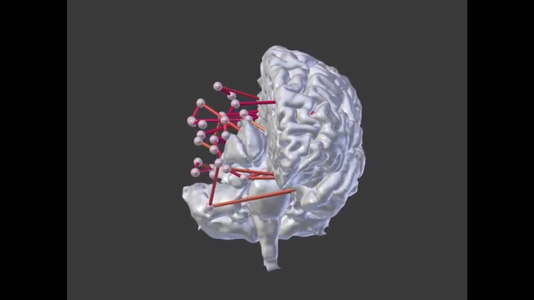
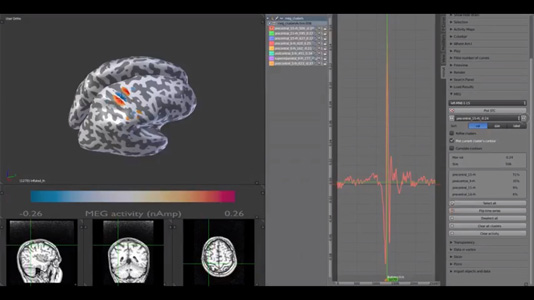
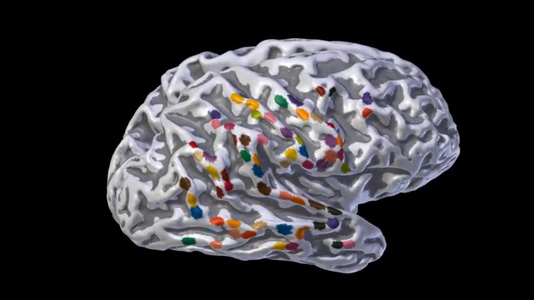
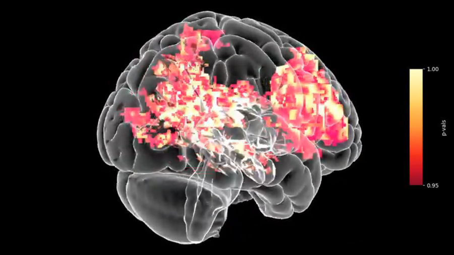
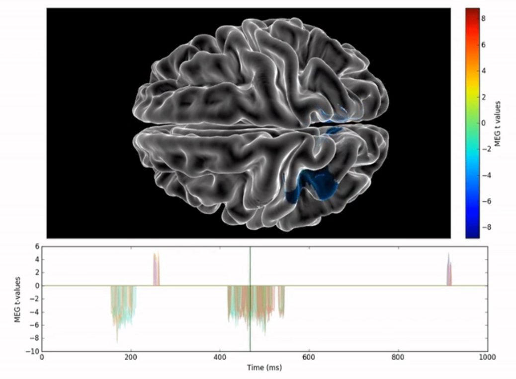
MMVT Gallery Figures Videos Figures AllAnatomyEEG MEGfMRI PETConnectivityDepth Electrodes Flat Brain MapFlatten brain with the DKT40 Freesurfer cortical atlas 3D slicer DKT40 AltlasRendering of the Freesurfer DKT40 cortical atlas Medial WallHiding one hemisphere for visualizing the medial wall Transparent Half BrainHiding one side of the brain DKT40 Medial Wall ParcellationThe medial wall parcellated using the DKT40 Freesurfer atlas Transparent BrainTransparent brain with the pial surface in the slices viewer Cortical CurvaturesPlotting the cortical curvatures using colors from the DKT40 Freesurfer atlas Skull ThiknessPanel for calculating the skull thickness and attaching a device DKT40 atlas in solid mode Zooming in the slices viewerUsing interpolation to zoom in the MRI slices viewer DKT40 Atlas ContoursDKT40 Freesurfer cortical atlas contours Depth ElectrodesDepth Electrodes in a rendered 3D slicer rendered brain (CT in blue) & evoked response DKT40 Atlas Contours on the Flat Brain Inflating the BrainSemi inflated brain . . . . . . . . . . . . . . . . . . . . . . . . . . . . . . . . . . . . . . . . . . Videos Play Video Depth Electrodes with Outer Skin Play Video Inflating & Rotating Brain Play Video MEG Helmet with Outer Skin Play Video Inflating Brain Inflating brain, from pial to inflated and to flat, with MEG evoked response. Play Video 3D Brain Slicing Play Video Inflating Brain Inflating brain, from pial to inflated and to flat, with MEG evoked response. Play Video ECOG Play Video Labels Connectivity Play Video Depth Electrodes Play Video EEG Helmet Play Video fMRI Resting State Connectivity and Raw Play Video Findig Electrodes in CT Play Video ECOG vs MEG Resting State Power-spectrum Cortical labels were created around the ECOG electrodes and their power-spectrum was calculated base on MNE source estimate. Play Video ECOG vs MEG Resting State ECOG LFPs vs MEG source estimate around each electrode using MNE. Play Video Skull Thikness Play Video Selecting Electrodes Play Video Streaming Play Video MMVT Presentation MMVT presentation to the director of Massachusetts General Hospital Epilepsy Service Dr. Andrew James Cole. Play Video fMRI Rest Connectivity Play Video MEG Clusters Play Video Inflating ECOG Electrodes We created a cortical label around each ECOG electrode and inflated the brain. See what happens to the grid shape. Play Video Volumetric Statistical Test Play Video Tempspatial ttest Results of MEG Source Estimate